Nuclei by layer
Layer boundaries are drawn roughly. They count every nucleus, not just neurons.
| Layer | Nuclei | Share |
|---|---|---|
| I | 7,871 | |
| II | 10,463 | |
| III | 14,203 | |
| IV | 4,200 | |
| V | 16,215 | |
| VI | 9,679 |
not yet releasedmouse mec
This dataset page is not public yet. Enter the secret access code to read it.
Seung Lab · Princeton
connectomic reconstructionmouse mec
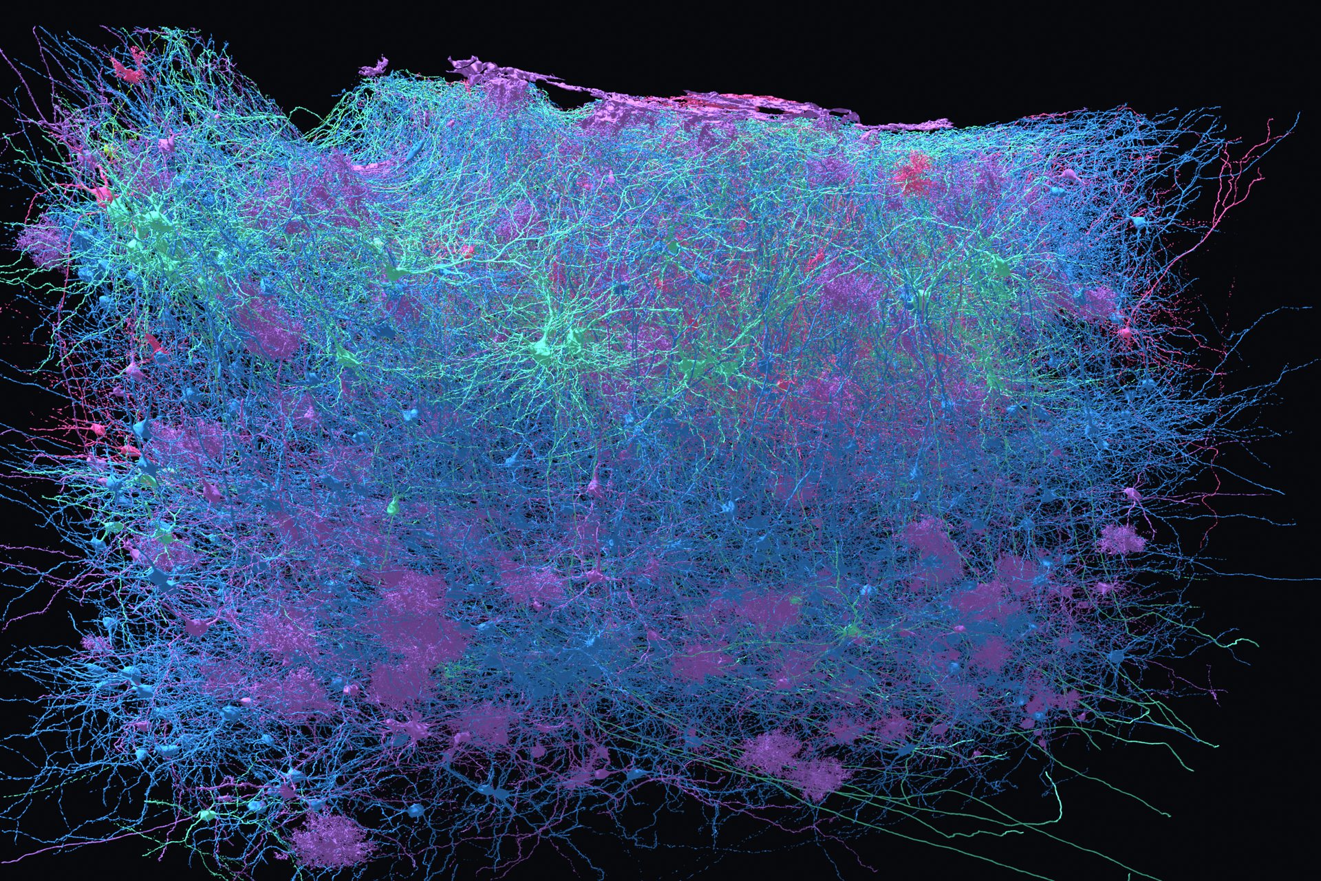
A part of the brain that helps keeps track of where you are. We imaged a block of it in electron microscopy, and now it needs mapping. It is the newest volume open to EyeWire citizen scientists in EyeWire II.
Put a mouse in a room and record a single cell in its medial entorhinal cortex, and something strange happens. The cell fires when the animal stands in one spot, then fires again a fixed distance away, and again, and again, until the firing positions trace a hexagonal grid across the whole floor. These are grid cells. Edvard and May‑Britt Moser found them in 2005, and shared the 2014 Nobel Prize in Physiology or Medicine with John O'Keefe for the work.
Nobody knows how the grid is built. The leading explanations disagree about what the wiring should look like, which is exactly the kind of argument a connectome can settle. The medial entorhinal cortex is also the main gateway from the cortex into the hippocampus, so its wiring shapes what the memory system receives in the first place. It is one of the first regions to degenerate in Alzheimer's disease.
The region sits at the back and underside of the cortex, in the retrohippocampal area, pressed up against the hippocampus it feeds. It is a thin sheet of cells folded over on itself, which is why a block of it a couple of millimetres across contains every cortical layer at once. You can see exactly where it is in the interactive mouse brain.
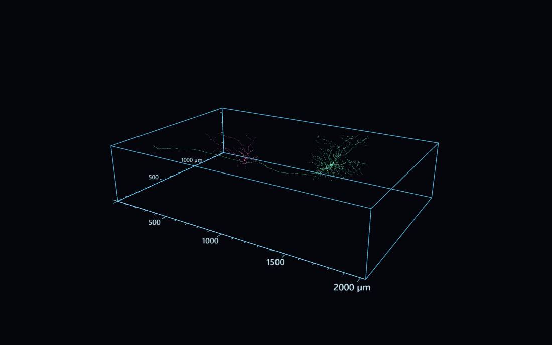
Two reconstructed cells inside the imaged volume, at true relative size and in the positions they actually occupy.
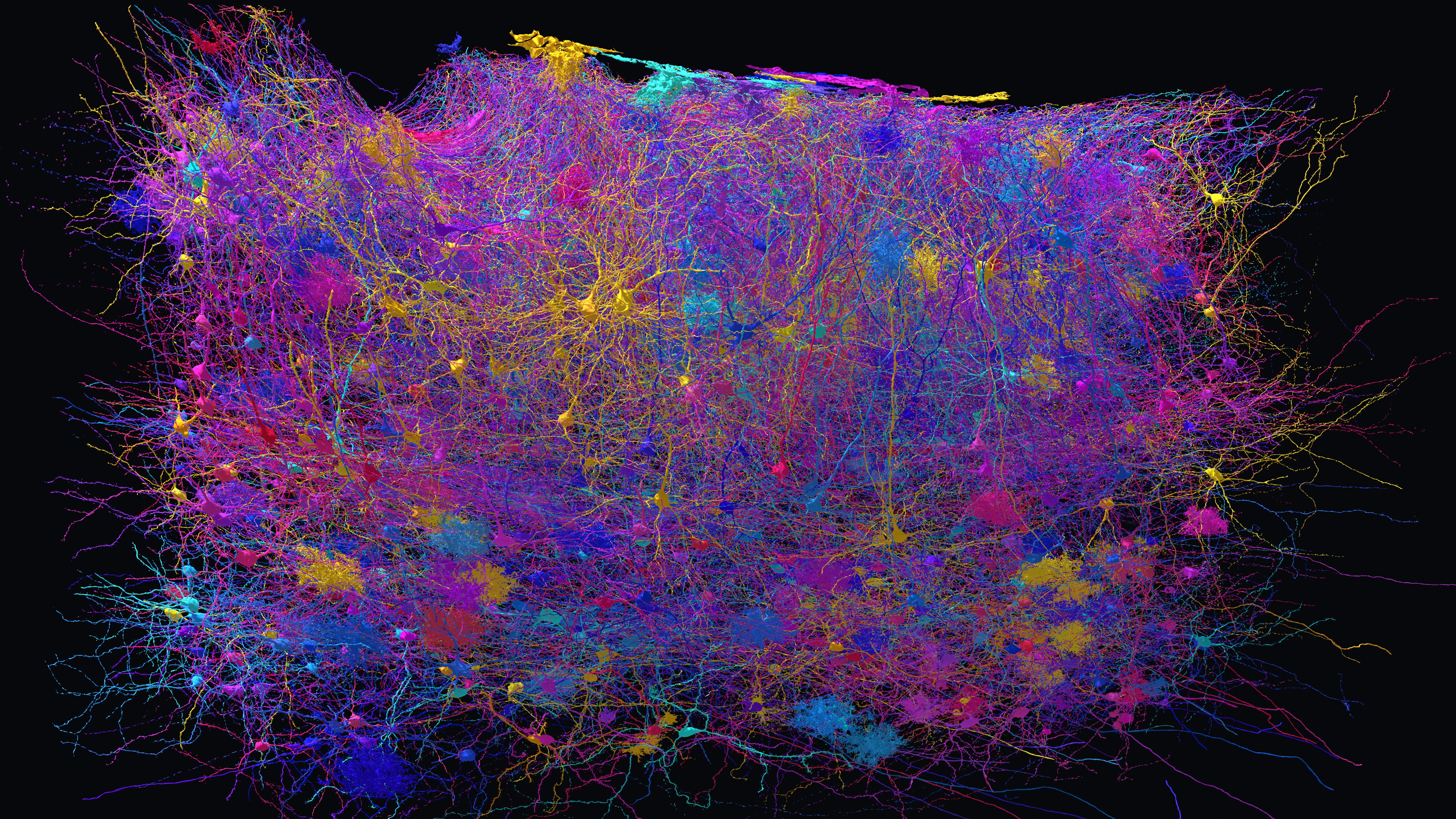
Every nucleus in the block has been marked with a point, assigned to a cortical layer, and sorted into a cluster by nucleus size. These are early numbers and will change as proofreading advances.
Of the 62,631 nuclei, 14,112 fall into the two clusters that carry a cell type name, pyramidal and stellate. The other 48,519 sit in six unnamed clusters. Some of those are neurons, interneurons especially, and some are glia, blood vessels and other non‑neuronal cells.
So the number of neurons in this block is at least 14,112 and certainly a good deal higher, and pinning it down is open work. The clustering is by nucleus size, which correlates with cell type but does not determine it, so every type on this page is a presumed type until a human has looked at the cell's shape.
Layer boundaries are drawn roughly. They count every nucleus, not just neurons.
| Layer | Nuclei | Share |
|---|---|---|
| I | 7,871 | |
| II | 10,463 | |
| III | 14,203 | |
| IV | 4,200 | |
| V | 16,215 | |
| VI | 9,679 |
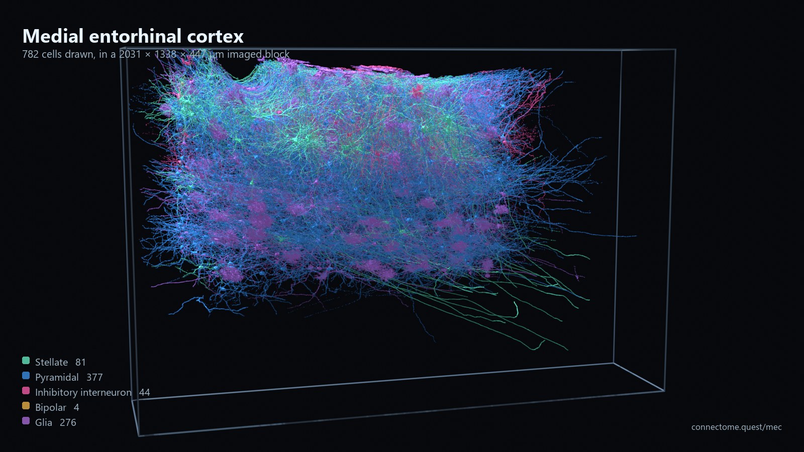
A sample of reconstructed cells, each drawn where it actually sits in the block and coloured by type. This is a sample, not a census: the block holds tens of thousands of cells and these are the ones picked out so far.
The three glial types are drawn as one colour here. The automatic type prediction calls glia by nucleus size, and when an expert checked two of them, both were wrong: the cell called an oligodendrocyte was an astrocyte, and the one called a microglia was an oligodendrocyte. Splitting them into three colours would be claiming something this project cannot yet support, so until they are proofread they are simply glia.
Here are some of the most common cell types found in the medial entorhinal cortex. We will be exploring them. Every one below is a real reconstruction from this block, and each card shows the segment ID so you can open it yourself.
Layer II
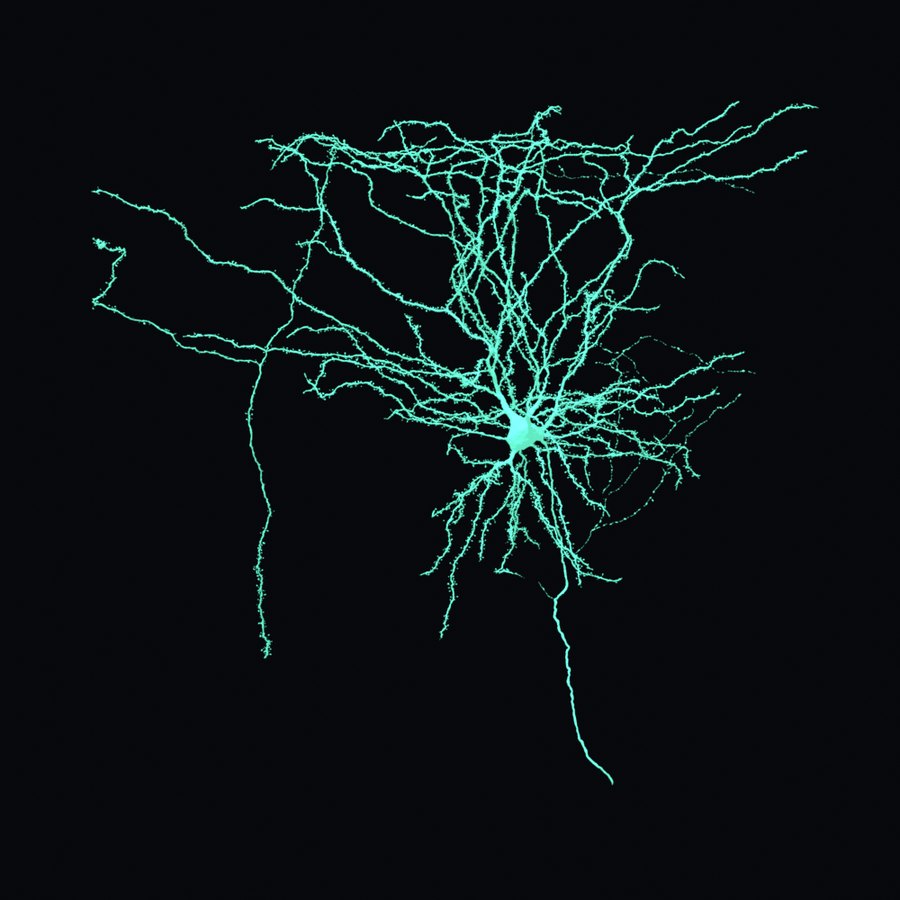
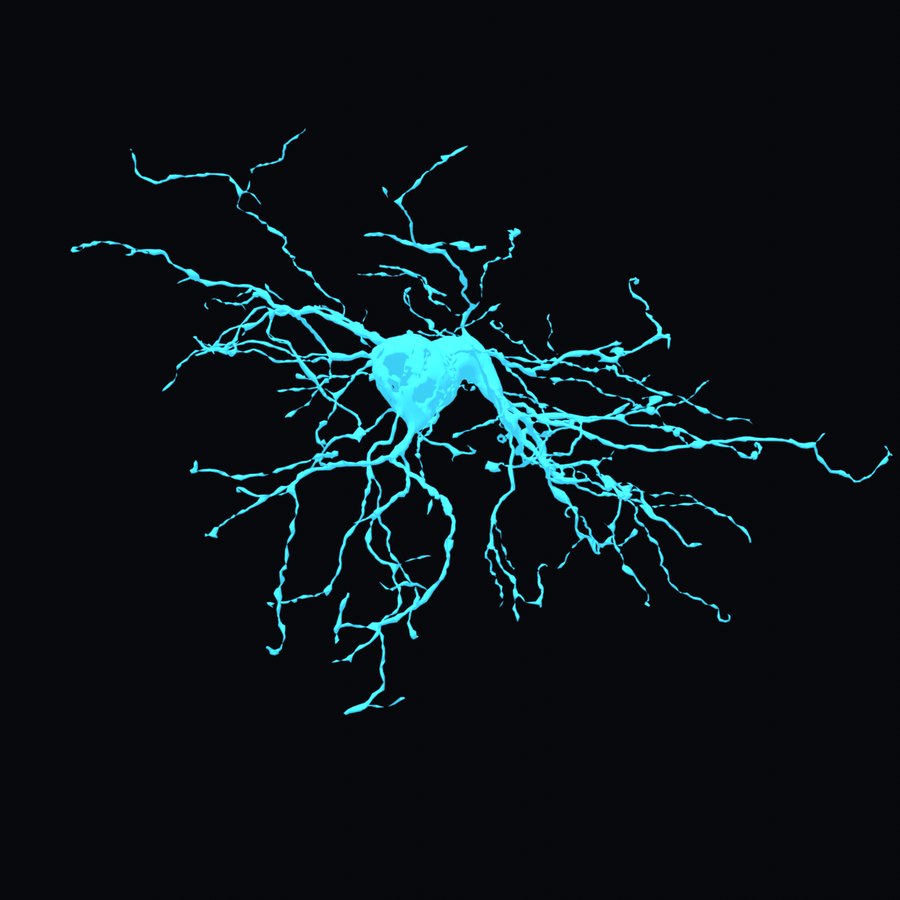
The grid cell candidate. Dendrites spread out from the soma in every direction, like a star, and the axon leaves for the hippocampus. Layer II of the medial entorhinal cortex is where grid firing was first recorded.
1,370 presumed in this block, 79% of them in layer II.
Layers II and III
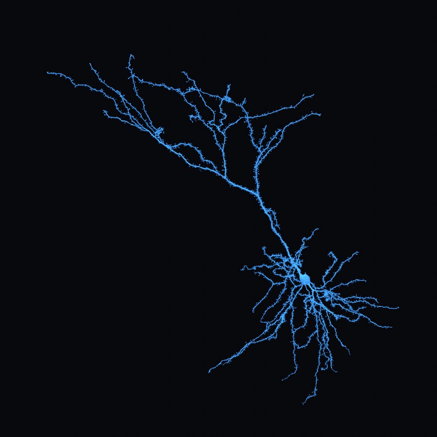
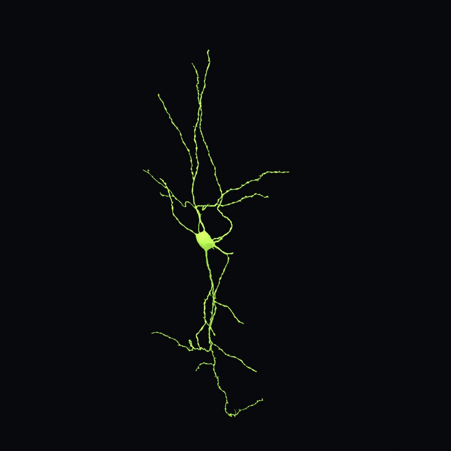
The other principal cell. One dominant dendrite rises from the top of the soma and a skirt of shorter ones spreads around its base. In the medial entorhinal cortex pyramidal and stellate cells grade into one another, and there is a real argument that they are a continuum rather than two types, so this one was chosen by eye as a clear example.
12,742 presumed in this block.
Layers II and III
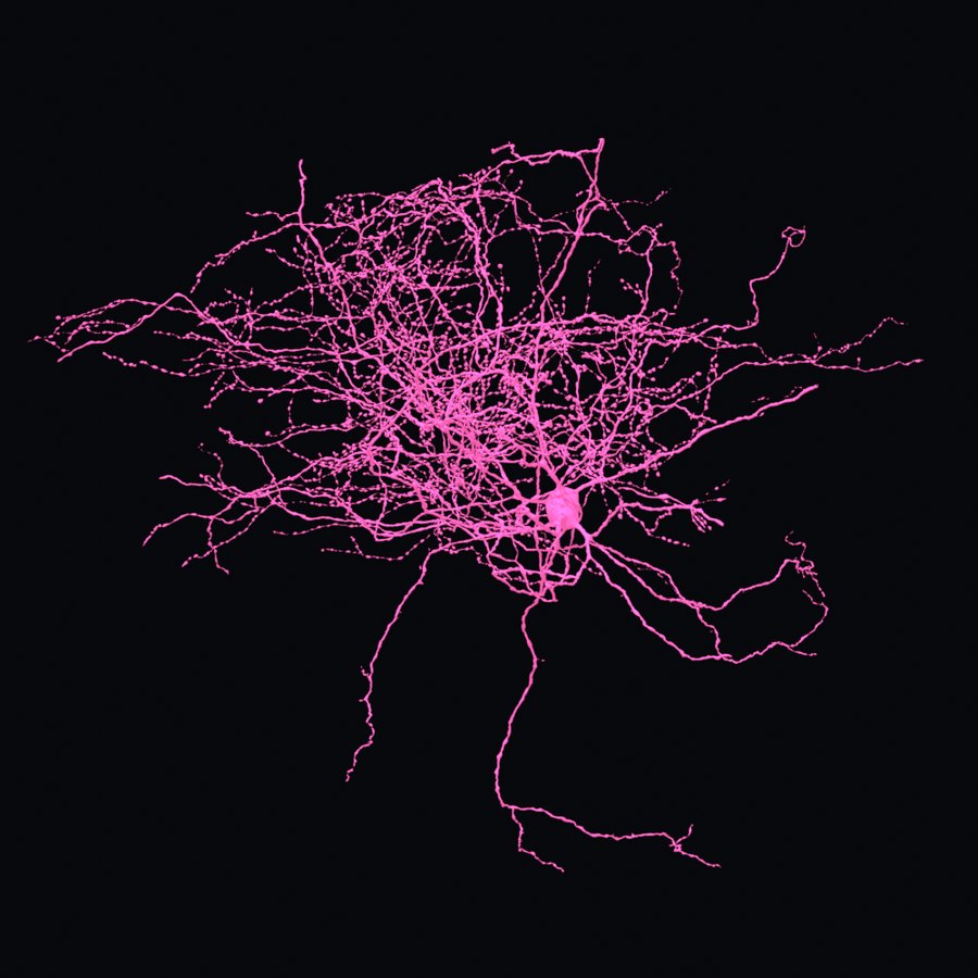
Local cells that quiet their neighbours, and the reason this volume needs people. They come in several shapes with no single look, and nucleus size does not separate them from pyramidal cells, so an automatic pass cannot find them. Human eyes can.
480 labelled so far. How many there are is not known.
Not a neuron
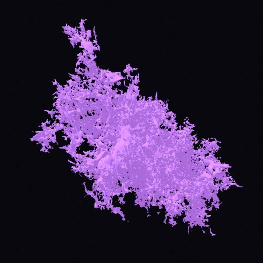
A star-shaped support cell whose fine processes wrap synapses and blood vessels. It is easy to mistake for a small neuron at a glance, which is part of why the neuron count in this block is still open.
188 labelled so far. This one is still an automatic call, and the glial predictions have already been wrong once.
Not a neuron
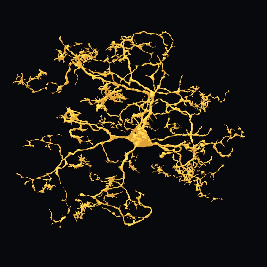
The cell that makes myelin. Follow the processes out and you will find small closed loops: each one is this cell wrapped right around a myelinated axon. A single oligodendrocyte insulates many axons at once.
137 labelled so far, but the automatic labels for glia are not reliable yet.
Not a neuron
The brain's resident immune cell. A small body with fine, heavily branched processes that are constantly reaching out and sampling the tissue around them. Cells like this are part of why the neuron count is still an open question.
24 labelled so far. Intact examples are rare, because microglia merge easily with the cells around them.
Layer II
Two processes, one out of each end of the cell body, and very little else. The least certain type on this page: only five have been labelled in the whole block, so there is almost nothing to check an example against.
5 labelled so far.
The cell type this region is known for, and the one most closely tied to grid firing. Here is where they sit and what is known about them in this block.
Stellate cells are the principal cells of layer II. Dendrites leave the soma in every direction rather than in one dominant trunk with a skirt beneath it, which is what separates them from pyramidal cells, and their axons carry the output of this cortex into the hippocampus. Grid firing was first recorded in layer II of the medial entorhinal cortex, and stellate cells are the population most often held responsible for it.
They are where the textbook says they should be. Of the 1,370 presumed stellate nuclei in this block, 1,085 are in layer II. That is 79%, and it comes out of a clustering by nucleus size that never saw the layer labels, so the layer and the type agree without either being told about the other.
| Layer | Stellate | Share |
|---|---|---|
| I | 42 | |
| II | 1,085 | |
| III | 223 | |
| IV | 8 | |
| V | 10 | |
| VI | 2 |
They avoid the pyramidal patches. Layer II is not a uniform sheet: pyramidal cells gather into islands and stellate cells occupy the water between them. Measured here, a stellate cell is 27 standard deviations less likely than chance to sit within 40 µm of a pyramidal cell, and that shortfall runs out to 271 µm. It is set out in Islands and ocean below.
What is still uncertain. "Stellate" here means a nucleus-size cluster, about 84% stellate where it has been checked against labelled cells. It is the cleanest of the automatic type calls on this page, and it is still a prediction rather than a cell anyone has looked at. The example in the gallery above is one we would particularly like proofread.
Layer II of this cortex is not a uniform sheet of cells. Pyramidal cells gather into patches, and stellate cells sit in the water between them. You can see it in the block before a single synapse has been traced, using nothing but the positions of the cell nuclei.
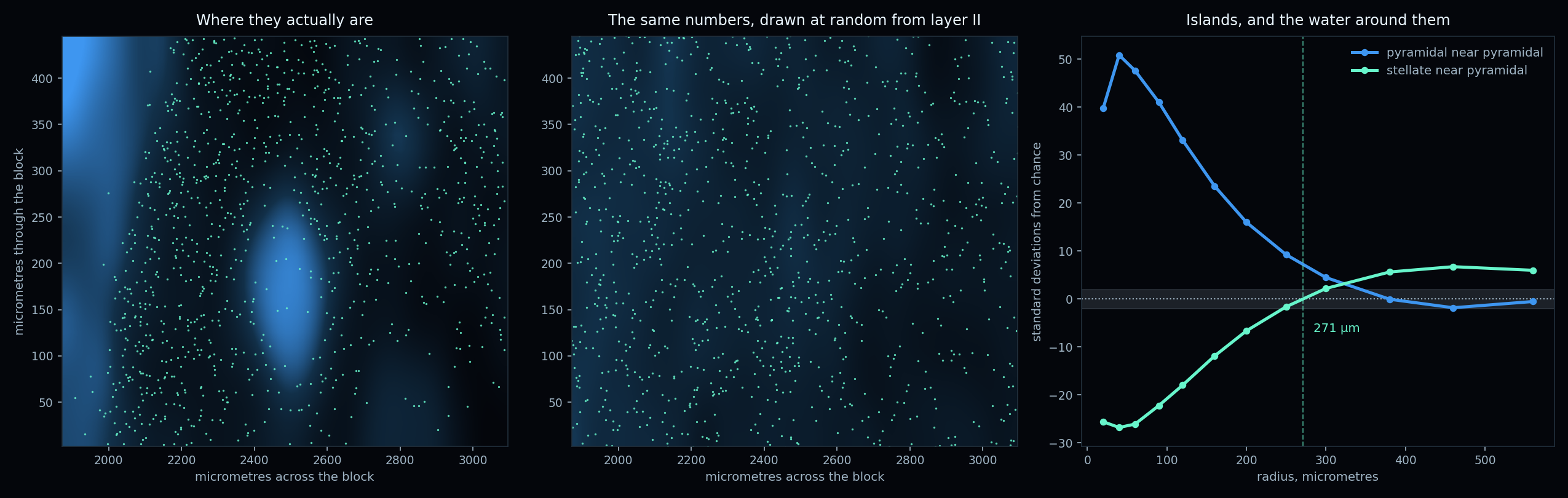
The control is the argument. Comparing against points scattered evenly in a box would find structure in any dataset, because layer II is a curved sheet inside a block and not a box. So every comparison here is against the same number of nuclei drawn at random from layer II itself, two hundred times over, which holds the sheet's shape and its density fixed.
Pyramidal cells sit near other pyramidal cells far more often than chance allows, 51 standard deviations above it at 40 µm. That excess then dies away and reaches zero by about 380 µm, which is what a patch of finite size looks like rather than a general tendency to clump.
Stellate cells do the opposite. Near a pyramidal cell there are far fewer of them than chance allows, 27 standard deviations below at 40 µm, and the shortfall persists out to 271 µm before turning into a mild excess beyond it. The two cell types are not merely clustered. They are arranged around each other.
None of this is new biology. Calbindin positive pyramidal cells in layer II of the medial entorhinal cortex were shown to sit in a patchy, grid like layout by Ray and colleagues in 2014, with reelin positive stellate cells occupying the space between the patches, and the arrangement was tied to grid activity by Naumann and colleagues in 2018. What is worth something here is that a reconstruction of this block recovers that layout independently, which is a good sign about the data. Zhihao Zheng spotted it in this volume straight away.
What this does not say. "Pyramidal" and "stellate" here mean nucleus size clusters, not cells anyone has looked at. The stellate cluster is about 84% stellate where it has been checked and sits almost entirely in layer II, where stellate cells belong. Mislabelled cells would blur an arrangement like this rather than create one, so the direction holds even though the labels are not exact. And none of it is about wiring yet. It says where cell bodies sit. The point of tracing the connectome is to find out whether the wiring respects it.
Open the volume in EyeWire II and start looking.
MEC is now in the dataset switcher in EyeWire II. Open the app, click Data in the top bar, and pick Medial Entorhinal Cortex. You will need to sign in with Google, twice, because the volume and the annotations live behind separate services.
Use a computer for this one. The phone build of EyeWire II does not carry MEC yet, so the button below opens the desktop app on purpose.
What works today: viewing the volume, loading cells, and proofreading them. If a reconstruction has two cells fused together you can split them, and if a branch has been cut off you can merge it back on. That is the core of the work.
What does not work yet: marking a cell complete, assigning a cell type, and anything that counts toward the leaderboard. Those write to annotation tables that have not been set up for this volume yet. We would rather tell you that plainly than have the buttons fail quietly. They are coming, and your proofreading in the meantime is real and is saved.
Everything on this page is rendered from the reconstruction itself. No cell here was drawn, straightened or stood in for by a model.
The volume was imaged, aligned and segmented by Eric Hammerschmith with Zhihao Zheng in the Seung Lab at Princeton. A team of professional proofreaders has been working through it ahead of this release. The reconstruction you see on this page came out of their work.
This page and the renders on it were made by Amy Sterling. Every image here is rendered from the reconstruction itself: no cell was drawn, straightened, or stood in for by a model.